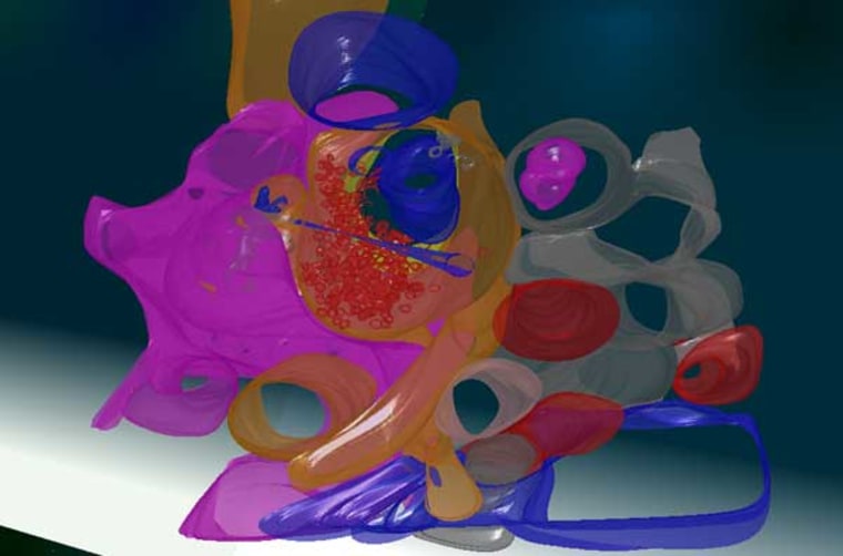
A new project aims to produce a Google Maps-like guide of the brain's labyrinthine structure. At a presentation here at the SIGGRAPH interactive technology and computer graphics conference, researchers highlighted how a complete 3-D model of the brain could spark a new era in neurological research.
Called The Whole Brain Catalog, the project compiles data from across the research spectrum, in a variety of forms. It takes MRI data, pictures of stained neurons and theoretical diagrams of brain circuitry and presents them in a way that scientists, doctors and 3-D animators can digest in a unified way. Those users then contribute back to the site, wiki-style, to produce an increasingly full model of the brain at every scale, down to the molecular level.
“My dream is that you can built these simulations at any level of resolution,” said Stephen Larson, a neuroscience researcher at the University of California, San Diego, who works on the Whole Brain Catalog. “The key is to have something smaller than the brain that is still big enough for simulations.”
Mapping out all the connections in the brain has proved one of the most difficult challenges facing neuroscientists today.
Indeed, they often compare the intricacy of the brain to the human genome — the name given to the entire library of human DNA — and have nicknamed the total map of all the brain’s links the “connectome”, Larson said.
Even before the Whole Brain Project is completed, scientists could use the data to run tests in the computer, instead of on live subjects. This would drastically increase the range of possible neurological experiments, while significantly cutting down the cost, time and variables of studies.
Ultimately, the connectome can only be understood through a complete 3-D model that maps out every molecule, neuron and wrinkle, Larson said.
“The brain is complex because it isn’t rectilinear like the world we live in, which makes it counter intuitive,” Larson said.
The Whole Brain Catalog remains a long way from finishing its task of mapping the connectome, and faces difficult challenges moving forward. For one, most of the data comes to the project in wildly different formats, and there is no way to automate the process of unifying the different types of input. Additionally, some regions of the brain receive far more research than others, leaving some areas mostly blank, Larson said.
However, that last problem also presents a new opportunity for the Whole Brain Project. Because the model reflects what data scientists generate, the model is as much a diagram of trends in neurological research as it is a map of the brain.
By looking at what areas are more or less defined in the Whole Brain Catalog model, scientists can determine what regions of the brain need more attention, and plan future research that crosses traditional boundaries that isolate different kinds of brain research, Larson said.
