Science News
Nikon Small World 2011

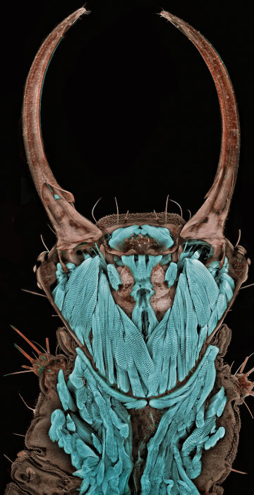
1st place: Portrait of a bug
Every year, the Nikon Small World Photomicrography Competition highlights the beauty and complexity of the world as seen through the light microscope. The top 20 pictures for 2011 focus on a wide range of biological and geological subjects, with a little fun thrown in. This 20x portrait of a green lacewing larva, captured using confocal microscopy, is the first-place winner. The scientist behind the image is Igor Siwanowicz of the Max Planck Institute of Neurobiology in Martinsried, Germany.
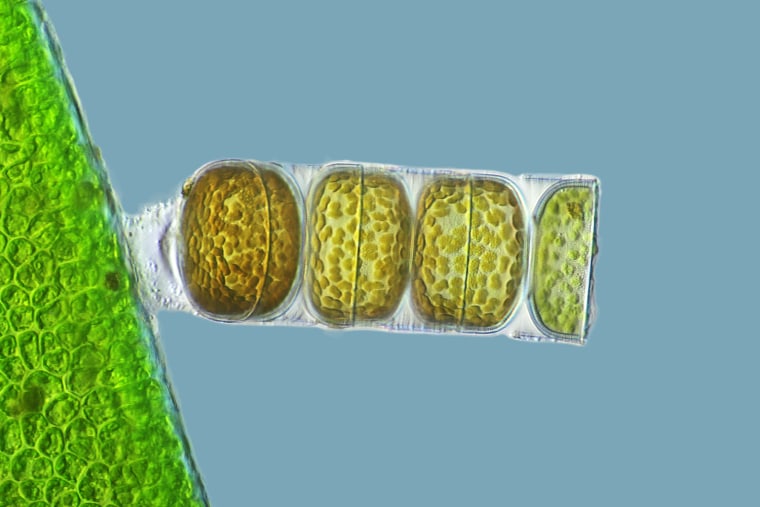
3rd place: It's alive!
Third place in the 2011 Nikon Small World competition goes to Frank Fox of the Fachhochschule Trier in Germany, for this 320x view of the colonial diatom known as Melosira moniliformis. These diatoms can chain themselves together to form a filamentous colony of cells, as seen here. The filaments are sometimes mistaken for seaweed. This image takes advantage of a microscope technique known as differential interference contrast.
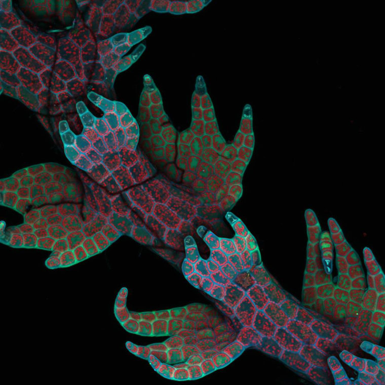
4th place: Lovely liverwort
Alien creatures? No, this 20x confocal microscopy image reveals the intrinsic fluorescence in a plant known as the "little hands" liverwort (Lepidozia reptans). The leaves of the tiny plant look like ... you guessed it, little hands. This image from University of British Columbia biologist Robin Young won fourth place in the Nikon Small World contest.

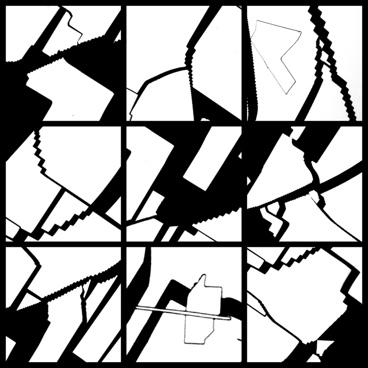
6th place: Black and white
This 50x brightfield image turns cracked gallium arsenide solar-cell films into a work of abstract art. The image, which won sixth place in the 2011 Nikon Small World contest, was submitted by Dennis Callahan, a solar-cell researcher at the California Institute of Technology.
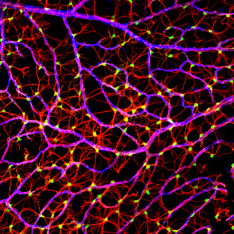
7th place: Some nerve!
Gabriel Luna, a researcher at the Neuroscience Research Institute at the University of California at Santa Barbara, used laser confocal scanning to produce this image of the nerve fiber layer in a mouse retina. The 40x image won seventh place in the 2011 Nikon Small World contest.
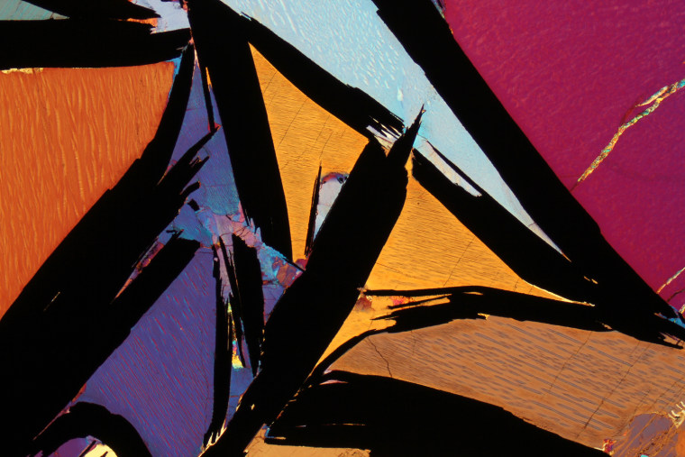
8th place: Graphic granulite
Polarized-light microscopy brings an artistic color scheme to this 2.5x image of graphite-bearing granulite from India's Kerala region. The picture was produced by Bernardo Cesare, a geologist at the University of Padua in Italy, and won eighth place in the 2011 Nikon Small World contest.
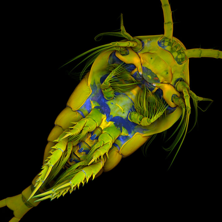
9th place: Micro-monster
This full-frontal view of a marine copepod (Temora longicornis) was created using confocal, autofluorescence and Congo red fluorescence microscopy. Copepods are tiny crustaceans, considered cousins of crawfish and water fleas. The 10x image was entered in the 2011 Nikon Small World contest by Jan Michels of the Christian-Albrechts-Universität zu Kiel in Germany, and won ninth place.
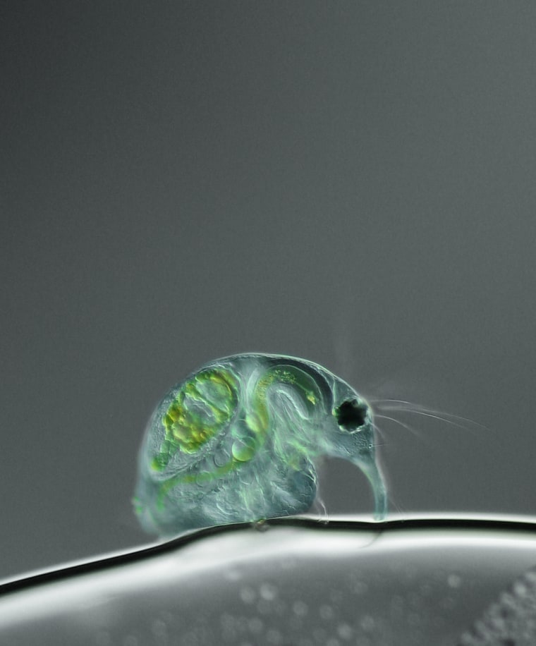
10th place: See-through flea
The innards of a freshwater water flea (Daphnia magna) become visible, thanks to differential interference contrast, in this 100x microscopic image. The picture, created by Joan Röhl of the Institute for Biochemistry and Biology in Potsdam, Germany, won 10th place in the 2011 Nikon Small World contest.
12th place: Immortal cells
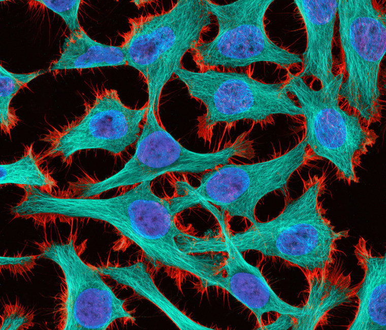
Two-photon fluorescence lends a glow to these HeLa cancer cells, an "immortal" cell line that gets its name from the donor, a Virginia-born woman named Henrietta Lacks. This 300x image, created by Thomas Deerinck of the National Center for Microscopy and Imaging Research in La Jolla, Calif., won 12th place in the Nikon Small World contest.
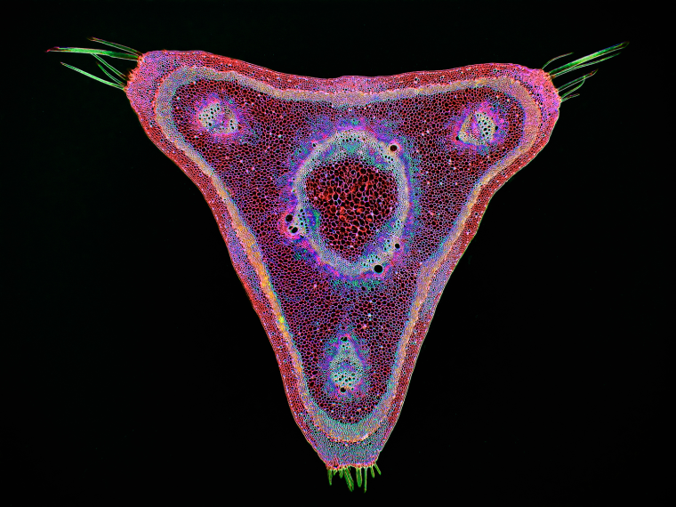
13th place: Pick your poison
This digitally inverted brightfield image shows a curare vine (Chondrodendron tomentosum) in cross-section. The vine is famous as a source for the poison used on South American arrows. The 45x image was entered by Stephen S. Nagy of Montana Diatoms in the 2011 Nikon Small World contest and won 13th place.
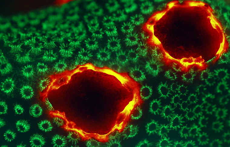
15th place: Coral on fire
This 12x image shows how a live specimen of lobe coral (Porites lobata) displays a pigmentation response with red fluorescence. The picture was made using epifluorescence microscopy with triple-band excitation. It earned 15th-place recognition in the Nikon Small World contest for James H. Nicholson of NOAA's Coral Culture and Collaborative Research Facility.
![[Improvision Data]
ImageName=Snapshot of Series008
TimeStampMicroSeconds=3382950489427790
TimeStamp=13:28:09,427 on 14 Mar 2011
ChannelName=
ChannelNo=1
TimepointName=1
TimepointNo=1
ZPlane=1
BlackPoint=0
WhitePoint=255
WhiteColour=255,255,255
XCalibrationMicrons=0,254
YCalibrationMicrons=0,254
ZCalibrationMicrons=1
TotalChannels=1
TotalTimepoints=1
TotalZPlanes=1
Software=Volocity 5.5.0
SampleUUID=51b4b6b6-7cc9-4916-bcfb-3d33fed30b3c](https://media-cldnry.s-nbcnews.com/image/upload/t_fit-760w,f_auto,q_auto:best/MSNBC/Components/Slideshows/_production/ss-111004-nikon-small-world/16 - Christopher Guerin.jpg)
16th place: Bionic cells
A confocal microscopic image, at 63x magnification, shows cultured cells growing on a bio-polymer scaffold. The picture was captured by Christopher Guérin, a researcher at the Flanders Institute of Biotechnology (VIB) in Ghent, Belgium. It earned 16th place in the Nikon Small World contest.
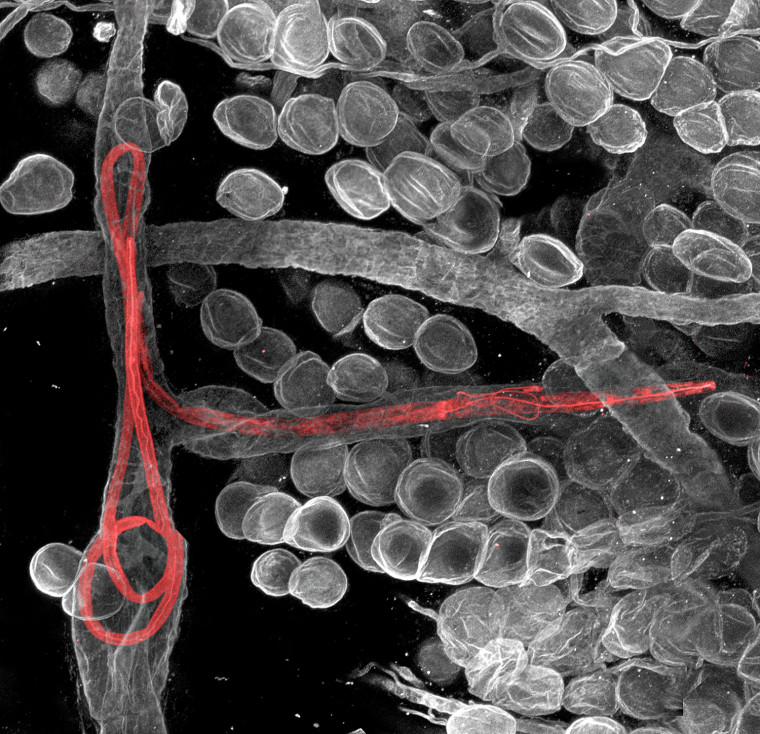
17th place: The worm turns
Filarial worms, highlighted in red, work their way through the lymphatic vessels in a mouse's ear. Witold Kilarski of Switzerland's EPFL-Laboratory of Lymphatic and Cancer Bioengineering captured this amazing 150x view using fluorescent confocal microscopy. It took 17th place in the Nikon Small World contest.
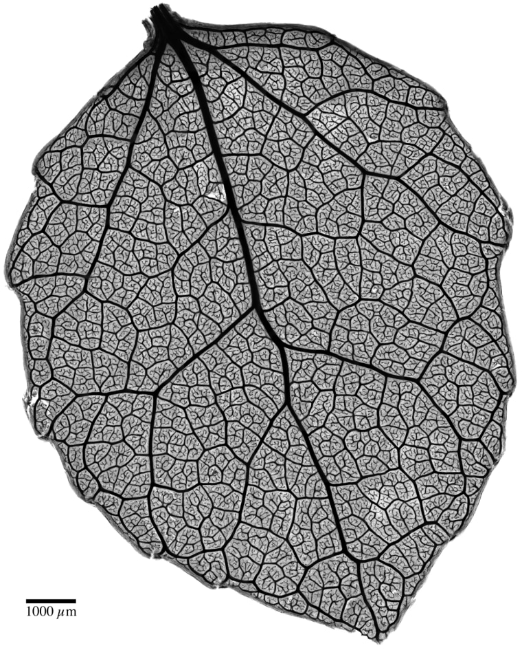
18th place: Inside a leaf
The network of veins in a quaking aspen leaf (Populus tremuloides) is highlighted in this 4x picture produced by Benjamin Blonder and David Elliott of the University of Arizona at Tucson. The brightfield image of safranin-stained tissue won 18th place in the Nikon Small World contest.
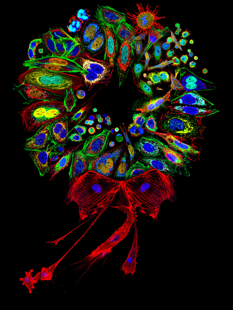
19th place: Season's proteins
A holiday wreath? Not exactly. This 200-2000x image, which took 19th place in the Nikon Small World contest, draws upon pictures of mammalian cells stained for various proteins and organelles. The single slice confocal cell mosaic was produced by the University of Pittsburgh's Donna Stolz.
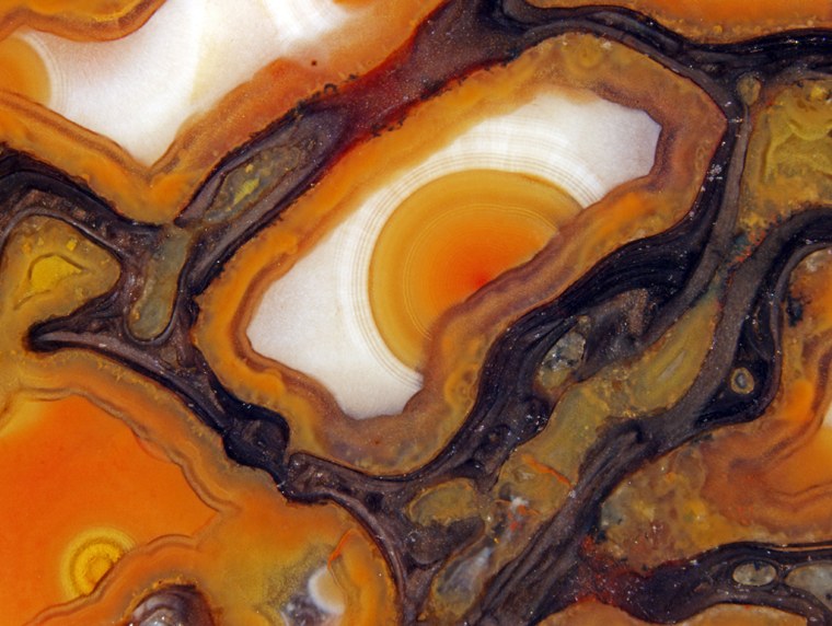
20th place: A dinosaur's stony stare
It almost looks as if a dinosaur's eye is glaring out from this picture. Actually, it's a 42x view of unpolished, agatized dinosaur bone cells, dating back about 150 million years. The picture was created by Douglas Moore of the University of Wisconsin at Stevens Point, using stereomicroscopy and fiber optics. It won 20th place in the 2011 Nikon Small World contest.