
Science News
Stunning scientific sights

Prize-winning eye
The International Science and Engineering Visualization Challenge honors artists and scientists who use visual media to promote understanding of scientific research. The annual competition is sponsored by the National Science Foundation and the journal Science. This picture, titled "Metabolomic Eye," is 2011's first-place winner in the photography category. It's a metabolic snapshot of the diversity of cells in a mouse eye retina, produced by neuroscientist Bryan William Jones of the University of Utah's Moran Eye Center using a technique called computational molecular phenotyping.
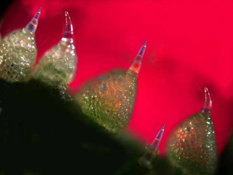
Hot as a cucumber
A colorful polarizing microscopic photograph of closely arranged trichomes on the outer skin of a cucumber was magnified 800 times by Robert Belliveau, a pathologist from Las Vegas. The image captures the sharp distal points of the trichomes, which are 40 times thinner than a sewing needle and perforate the mouthparts of herbivores. The globular base contains toxic, bitter cucurbiticins, the most bitter substances known, which can be detected by humans when diluted to one part in a billion. The image won honorable mention in the 2011 International Science and Engineering Visualization Challenge.

Microscopic cliff
When titanium aluminum carbide is placed in hydrofluoric acid, layers of aluminum are selectively etched away, resulting in two-dimensional layers of carbide weakly bonded to each other. This image shows a number of these layers. Like graphene, the individual layers can be isolated and their properties explored. This new family of layered solids is called MXene. The "People's Choice" photograph was created by Babak Anasori, Michael Naguib, Yury Gogotsi and Michel W. Barsoum of Drexel University.
![Tumor death-cell receptors on breast cancer cell
This illustration shows tumor death-cell receptors (DR5) on breast cancer cell surfaces targeted by the monoclonal antibody TRA-8, which was developed at the University of Alabama, Birmingham School of Medicine.
[Image courtesy of Emiko Paul and Quade Paul, Echo Medical Media; Ron Gamble, UAB Insight]
--](https://media-cldnry.s-nbcnews.com/image/upload/t_fit-760w,f_auto,q_auto:best/MSNBC/Components/Slideshows/_production/ss-120201-visualizations/120202-viz-04-tumor.jpg)
Battling a tumor
This illustration shows tumor death-cell receptors (DR5) on breast cancer cell surfaces targeted by the monoclonal antibody TRA-8, shown here in green. The anti-cancer technique was developed at the University of Alabama, Birmingham School of Medicine. Honorable mention, image courtesy of Emiko Paul and Quade Paul, Echo Medical Media; Ron Gamble, UAB Insight.
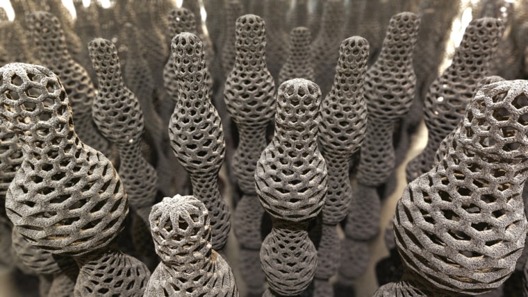
Nanotubes on parade
This 3-D illustration shows the production of variable-diameter carbon nanotubes.Yongteng Lu, an electrical engineering professor at the University of Nebraska-Lincoln, discovered laser-based production techniques that can precisely control the length, diameter and properties of carbon nanotubes. The illustration was developed to help Lu's team visualize nanoscale discoveries for diverse audiences. Honorable mention, image courtesy of Joel Brehm, University of Nebraska-Lincoln Office of Research and Economic Development.
![Exploring Complex Functions using Domain Coloring
Complex functions are of fundamental importance in many areas of mathematics, physics and engineering. The picture shows the visualization of a complex function using a specifically designed color scheme. Following a technique called 'domain coloring,' the color scheme assigns a certain color to every complex number, inducing a coloring of the function domain according to its values at every point. The picture allows to easily explore properties of the function, for instance, critical values such as zeros (black spots) or singularities (white spots). Contour lines indicate how the function deforms the complex plane. This modern visualization technique gives unprecedented insight into complex functions.
[Image courtesy of Konrad Polthier and Konstantin Poelke, Free University of Berlin]](https://media-cldnry.s-nbcnews.com/image/upload/t_fit-760w,f_auto,q_auto:best/MSNBC/Components/Slideshows/_production/ss-120201-visualizations/120202-viz-06-domain.jpg)
Delightful domains
Complex functions are of fundamental importance in many areas of mathematics, physics and engineering. This illustration shows the visualization of a complex function using a specially designed color scheme. Following a technique called "domain coloring," the color scheme assigns a certain color to every complex number, inducing a coloring of the function domain according to its values at every point. Honorable mention, image courtesy of Konrad Polthier and Konstantin Poelke, Free University of Berlin.
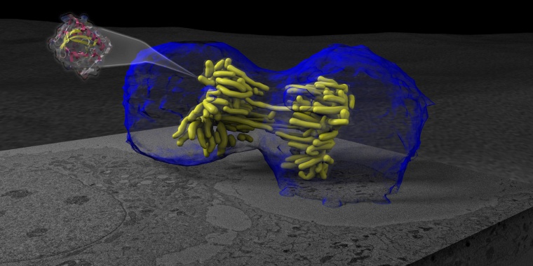
Separation of a cell
This illustration shows a cell undergoing mitosis or "cell division." The cell membrane is shown in blue, and the cell's chromosomes are shown in yellow. Mitosis is a well-studied and well-imaged phenomenon in two-dimensional images, but it's never before been seen quite like this. What makes this image special is the use of a new fluorescent protein called MiniSOG, shown flying out of the cell. People's Choice, image courtesy of Andrew Noske and Thomas Deerinck (National Center for Microscopy and Imaging Research, University of California, San Diego); Horng Ou and Clodagh O'Shea (Salk Institute).
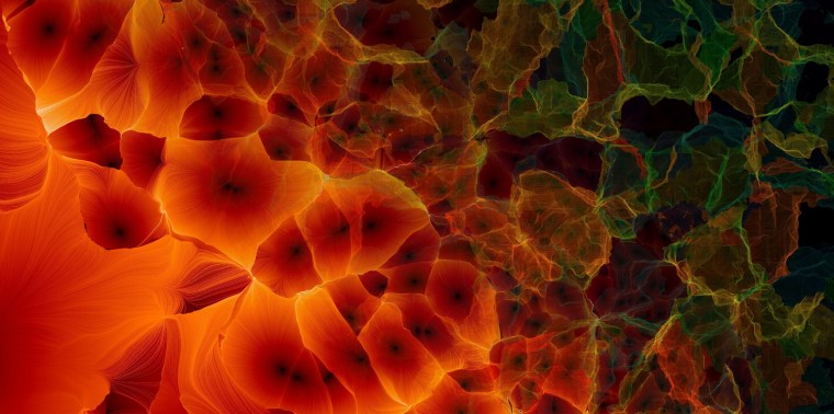
The cosmic web
This is just one piece of a poster showing different aspects of the cosmic web, the structure of voids and matter concentrations that make up our universe. The full poster shows the evolution of the cosmic web over billions of years. First-place, informational posters and graphics; image courtesy of Miguel Angel Aragon-Calvo (Johns Hopkins University), Julieta Aguilera and Mark SubbaRao (Adler Planetarium).
![The Ebola Virus
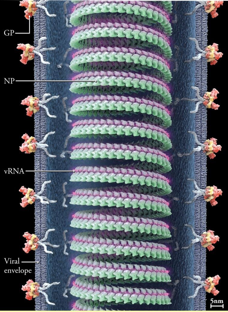
The Ebola Virus poster. View of the virion surface and of the ebola particle internal content.
[Image courtesy of Ivan Konstantinov, Yury Stefanov, Alexander Kovalevsky, Anastasya Bakulina; Visual Science]](https://media-cldnry.s-nbcnews.com/image/upload/t_fit-760w,f_auto,q_auto:best/MSNBC/Components/Slideshows/_production/ss-120201-visualizations/120202-viz-09-ebolaposter.jpg)
The Ebola virus
An eye-catching poster provides a three-dimensional, annotated view of the Ebola virus' supramolecular structure. Honorable mention, informational posters and graphics; image courtesy of Ivan Konstantinov, Yury Stefanov, Alexander Kovalevsky and Anastasya Bakulina of Visual Science.
Electron microscopy
Visualization is an essential research component when trying to understand the complex pathogenesis of infectious diseases. This illustration is taken from a poster developed to show how transmission electron microscopy is used to study virus particles. People's Choice, informational posters and graphics; image courtesy of Fabian de Kok-Mercado, Victoria Wahl-Jensen and Laura Bollinger of the National Institute of Allergy and Infectious Diseases IRF.
Foldit
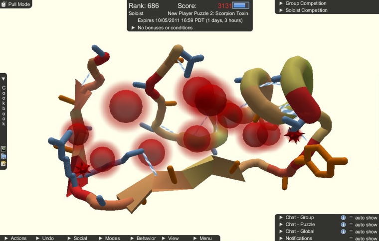
Foldit is a game designed to tackle the problem of protein folding. By learning more about the 3-D structure of proteins (or how they "fold"), scientists can better understand their function, and get a better idea how to combat diseases, create vaccines and even find novel biofuels. In Foldit, players are presented with a model of a protein, which they can fold by using a variety of provided tools. The game evaluates how well the player has folded the protein and gives them a score. Scores are uploaded to a leaderboard, allowing for competition between players from all around the world. First place, interactive games; screenshot courtesy of Seth Cooper, David Baker, Zoran Popović, Firas Khatib, Jeff Flatten, Kefan Xu, Dun-Yu Hsiao and Riley Adams, Center for Game Science at the University of Washington.
Velu the Welder
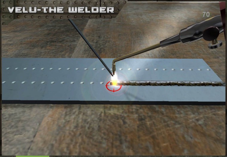
Welding is a method used for binding metal or nonmetal structures. In this game, an apprentice by the name of Velu gets basic training in the art of welding. The game is designed to help the player get acquainted with the craft of welding. Developed as a technology demonstrator for the National Skills Development of India, this game will be used to train millions of apprentices in a cost-effective way. People's Choice, interactive games; screenshot courtesy of Muralitharan Vengadasalam, Ganesh Venkat, Vignesh Palanimuthu, Fabian Herrera and Ashok Maharaja of Tata Consultancy Services.
Meta!Blast 3D
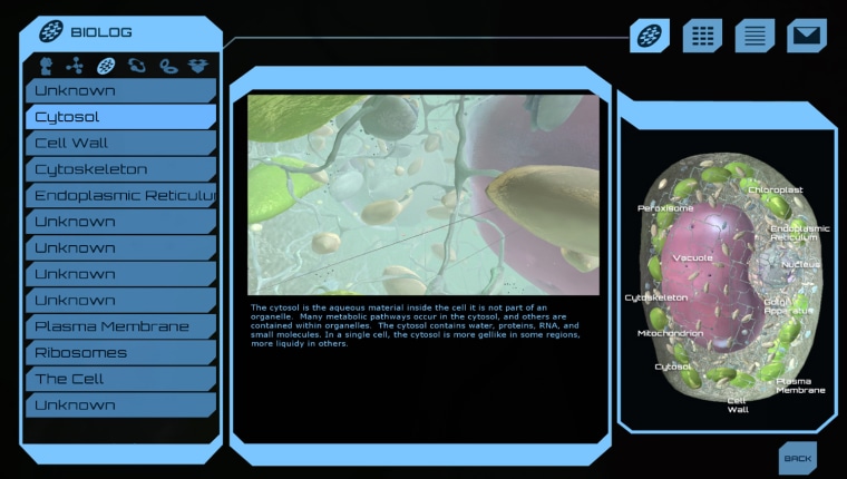
The year is 2052. An unknown pathogen is decimating Earth's vegetation. An accident has stranded a team of scientists inside a photosynthetic cell. You, the lab dishwasher they left behind, are their only hope. Can you navigate the bioship through microscopic hazards, solve metabolic puzzles, and re-engineer microorganisms to save the planet? Meta!Blast communicates biological concepts to high-school students. Honorable mention, interactive games; screenshot courtesy of W. Schneller, P.J. Campbell, M. Stenerson, D. Bassham, and E.S. Wurtele of Iowa State University.
![Powers of Minus Ten
Powers of Minus Ten (POMT) was originally conceived as an iPad app that would allow the user to zoom into the human body, exploring worlds at different levels of magnification (e.g. tissue, cellular, molecular, subatomic). In this version of POMT, the user is able to zoom into the human hand down to the molecular level. Three cell types and a variety of other structures can be viewed and explored. Users can also investigate structures in the ÔLabÕ area of the app, and review what they have discovered via timed mini-games. The app covers basic topics in biology such as the Phases of Mitosis and DNA Replication. POMT is available for iPads, iPhones, PCs, Macs, and as a web-based game. Future versions of POMT will allow the user to explore different subjects such as plants, minerals, water droplets, etc, as well as explore ÔdeeperÕ at the atomic and subatomic levels of magnification.
[Image courtesy of Laura Lynn Gonzalez; Green-Eye Visualization]
--](https://media-cldnry.s-nbcnews.com/image/upload/t_fit-760w,f_auto,q_auto:best/MSNBC/Components/Slideshows/_production/ss-120201-visualizations/120202-viz-14-powers.jpg)
Powers of Minus Ten
Powers of Minus Ten was originally conceived as an iPad app that would allow the user to zoom into the human body, exploring worlds at different levels of magnification. In this version of POMT, the user is able to zoom into the human hand down to the molecular level. Three cell types and a variety of other structures can be viewed and explored. POMT is available for iPads, iPhones, PCs, Macs and as a Web-based game. Honorable mention, interactive games; screenshot courtesy of Laura Lynn Gonzalez, Green-Eye Visualization.
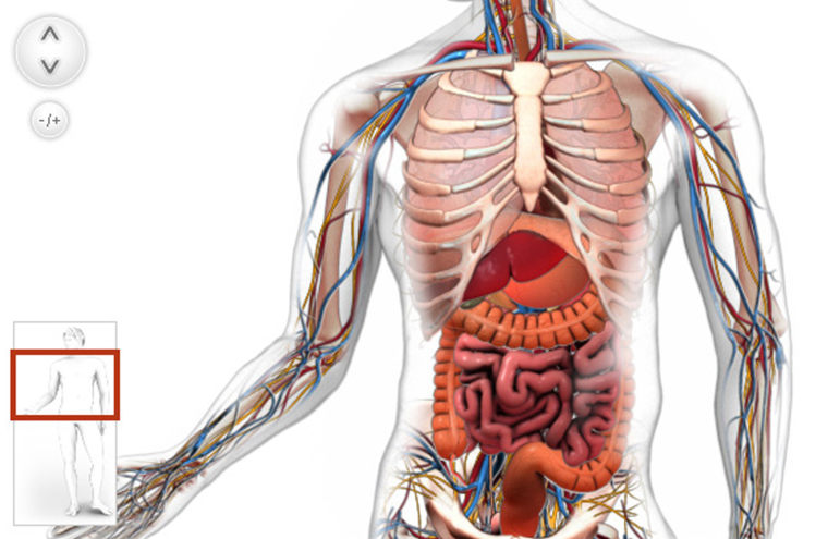
Build-a-Body
Put on your surgical gloves and get ready for the virtual operating room: You can learn about the organs of the human body with a drag-and-drop game called Build-a-Body. Choose organs from the organ tray and place them in their correct position within the body to create organ systems. Honorable mention, interactive games; screenshot courtesy of Jeremy Friedberg, Nicole Husain, Ian Wood, Genevieve Brydson, Wensi Sheng, Lorraine Trecroce, Kariane St-Denis, David Rowe, Ruby Pajares, Arij Al Chawaf, Shaun Rana and Nancy Reilly of Spongelab Interactive.