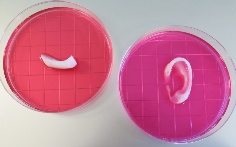
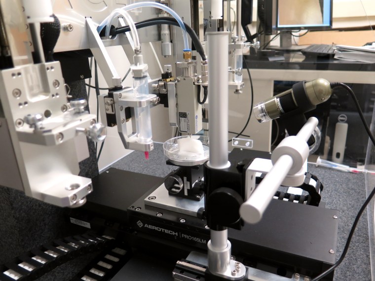
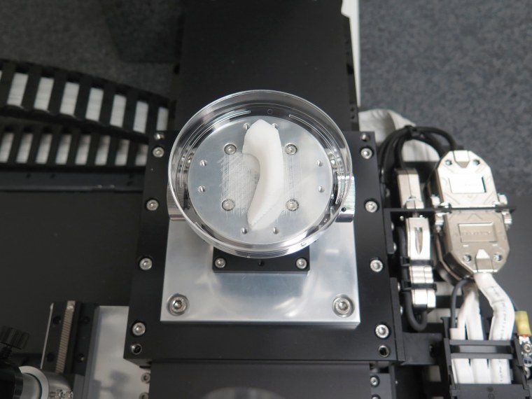
A team at Wake Forest University has used a combination of living cells and a special gel to print out living human body parts — including ears, muscles and jawbones.
It’s an advance on previous attempts, which either involved making a plastic scaffold and then trying to get cells to grow in and on it, or that printed out organ shapes that ended up being too floppy and dying.

The new approach mixes live cells with a gel that starts out as a liquid but quickly hardens to the consistency of living tissue, and layers them in with tiny tunnels that serve as passages for nutrients to feed the cells until blood vessels can grow in and do the job naturally.
“We are actually printing the scaffolds and the cells together,” said Dr. Anthony Atala of the Wake Forest University Institute for Regenerative Medicine, who led the study team.
“We show that we can grow muscle. We make ears the size of baby ears. We make jawbones the size of human jawbones. We are printing all kinds of things,” Atala told NBC News.
Writing in the journal Nature Biotechnology, the team describes both the new 'bioprinting' technology and the organs they have been able to grow using it.
“We present an integrated tissue–organ printer (ITOP) that can fabricate stable, human-scale tissue constructs of any shape,” they wrote.
“The correct shape of a tissue construct is obtained from a human body by processing computed tomography (CT) or magnetic resonance imaging (MRI) data in computer-aided design software.”
Atala has been working for more than a decade to make grow-your-own organ transplants. He's not the only one trying — other teams are working on bioprinted hands, for instance.
In 2006, Atala's team made the first full organ ever grown and implanted into a human — the bladder — and rabbit penises that were the first solid organs.
He’s been working under contract with the Armed Forces Institute of Regenerative Medicine, which is seeking new ways to help military personnel injured in battle. But the principles could apply to any patient needing a new ear, organ or a replacement for a shattered jaw.
The challenge is that living body parts are complicated. It’s not enough to make, say, a heart-shaped blob of tissue. Even a simple structure, like an ear, has several types of cells and they must all be fed by tiny capillaries.
“What happens if you don’t have them is the surface gets fed but if you make them any larger the central core with not get fed and they will die,” Atala said.
Atala’s team started out with actual inkjet printers but they’ve now developed customized devices.
“We started working with the printers about 12 years ago, trying to design printers specific to human tissues,” he said. “Nature itself gives us this limit where you cannot really print tissues in volumes larger than 200 microns.”
Anything thicker and the cells on the inside die. “Because the cells need vital nutrients. as we print the cells we can create microchannels. It’s like a highway,” he said.

And within 24 hours, blood vessels start to sprout in these microchannels, Atala said.
A second challenge is getting the bioprinted material to take on the right consistency at the right time.
“You want the cells to go through the nozzle as a liquid. But you don’t want it to land as a liquid because it will just make a blotch. You want it to land like a gelatin,” Atala said.
“Then as it hardens you want about as hard as a gummy bear.”
The result is a mixture of polymers, living cells and the nutrients they need. “We call it bioink,” Atala said.
The team’s also working to print out livers, lung tissue and kidney tissue.

It’s still years away from being used in actual human patients, but what Atala hopes for is a way for people to get custom-made transplants using their own cells, or closely matched cells, grown to just the right size and shape.
Currently, people must rely on organs taken from other peoples who have just died, or a kidney or piece of liver from generous and courageous live donor willing to go through the surgery.
Recipients must then take drugs for the rest of their lives to make their bodies tolerate the foreign tissue.
According to the United Network for Organ Sharing, more than 121,000 Americans are on the waiting list for an organ.
