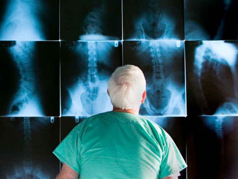
Although it’s relatively rare, mix-ups of patients’ X-rays can lead to dire consequences.
One way to get around the problem of such “wrong patient” errors is to take a photo of the patient’s face at the same time the X-ray is shot, a new study suggests.
If a doctor is looking at the wrong X-ray, the fallout could be catastrophic, said Dr. Srini Tridandapani, the author of the study presented at the annual meeting of the American Roentgen Ray Society.
“The patient could be diagnosed with cancer and then get an operation he shouldn’t have while the patient who should have gotten the cancer diagnosis isn’t getting the surgery. So you could be affecting two patients,” said Tridandapani, an assistant professor of radiology and imaging sciences at Emory University.
No one knows exactly how many times patients are matched with the wrong X-ray each year, said Tridandapani, who conducted the study jointly with the Georgia Institute of Technology. But it’s estimated that these kinds of identification errors occur in 1 out of every 10,000 patients.
While that may seem like a small number, companies like Motorola aim to have no more than one chip in a million fail, Tridandapani noted.
“I think human beings are more precious than chips, so I don’t accept a rate of 1 in 10,000,” he added. “I think we need to get beyond 1 in a million.”
Tridandapani came up with the idea of adding patient photos to X-rays after he answered a call and the image of the caller appeared on his phone.
“It occurred to me that we should be adding a photograph to every medical imaging study,” he said.
Errors could be reduced simply by adding photos to patient X-rays, he thought. To test his theory, Tridandapani rounded up 200 pairs of X-rays (one pair per patient) that he gave to 10 radiologists to read.
Each radiologist got 20 pairs of X-rays. Each set of X-rays contained a few pairs that were actually from different patients. The first time he ran his experiment, the radiologists got only the X-rays. The second time, the X-rays came with photos of the patients.
When there was no photo, the group of radiologists caught only three out of 24 mismatches – about 13 percent. When photos were included with the X-rays, they caught 16 out of 25 errors – or 64 percent.
Part of the problem is that doctors don’t expect to get the wrong X-ray and they often don’t recognize the mistake when it happens, Tridandapani said. They might try to explain away disparities instead of recognizing the errors.
Dr. Albert Wu, who has studied near-misses in medicine, said the new method might, indeed, avoid some dangerous mix-ups.
“This study, on its face makes a lot of sense to me,” said Wu, a professor of health policy and management at the Johns Hopkins Bloomberg School of Public Health and an attending physician at the Johns Hopkins Hospital.
“If you have the wrong films for someone about to have a procedure you could have a terrible result,” Wu said.
Still, Wu said, there are possible downsides. When you add photos, radiologists might take longer to read X-rays. And there’s also a question of privacy -- people might not want photos of themselves included in their medical files. Overall, he said, the added safety probably outweighs these concerns.
“I think it’s a pretty neat way to improve patient safety,” said Dr. Ashish Jha, a professor of health policy at the Harvard School of Public Health. “I see very little downside to it, other than the possibility that a physician might read the X-rays differently if it was a man or a woman -- and I think in general that’s unlikely.”
Even though Dr. Mitchell Schnall believes that the new method makes sense, he’s not sure that his colleagues will jump at the chance to implement it.
“There’s a lot of pressure on radiology in terms of efficiency,” said Schnall, a professor and chair of the department of radiology at the University of Pennsylvania. “Anything that adds to the workload gets looked at with some skepticism.”
Schnall said radiologists will worry that the addition of photos will mean that each case will take longer to analyze. But, he added, the photos might actually speed things up.
“For example,” Schnall said, “when you’ve got a patient in the ICU setting there are many tubes and wires in the patient, an external photo would help us know whether those are internal or external.”
Related stories:
