IE 11 is not supported. For an optimal experience visit our site on another browser.
Dr. Alvaro Esteves Migotto
Universidade de São Paulo, Centro de Biologia Marinha
São Paulo, São Paulo, Brazil
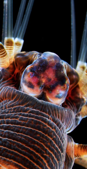
Marine worm
Stereomicroscopy, Darkfield
20X

Science News

Nikon Small World 2013

See a worm gone wild, a twisty piece of plankton and other top-20 photos from the 2013 Nikon Small World Photomicrography Competition.
/ 20 PHOTOS
Dr. James Burchfield
The Garvan Institute
Sydney, New South Wales, Australia 
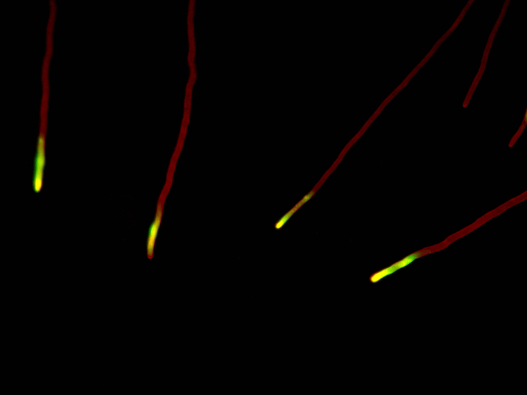
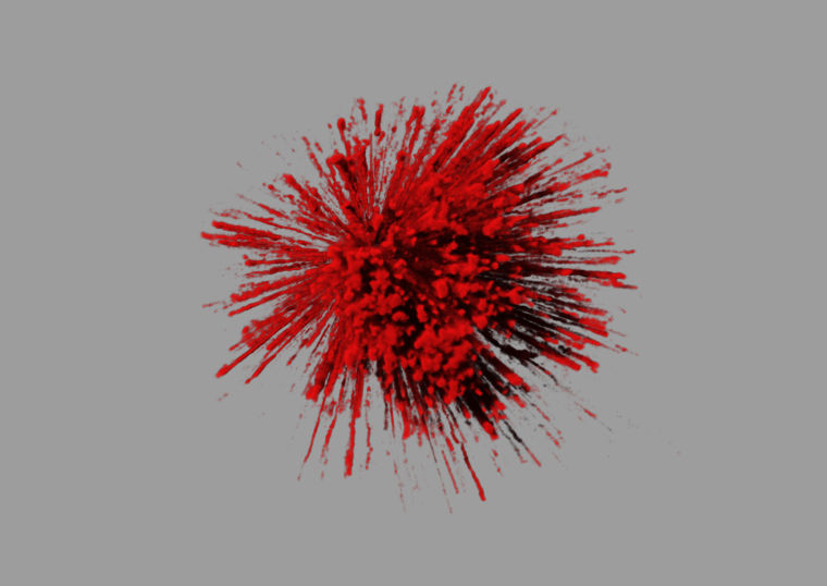
The explosive dynamics of sugar transport in fat cells
Live Cell Total Internal Reflection Fluorescence
1,000,000X

Countdown for a small world

The Nikon Small World Photomicrography Competition has been celebrating the wonders of microscopic realms since 1974. The judges for the 2013 contest sifted through more than 2,000 entries to come up with their top 20. This 20th-place entry was submitted by James Burchfield of Australia's Garvan Institute. It shows the explosive dynamics of sugar transport in fat cells. Browse through our slideshow to count down to the top-rated image.
Imaris X64 6.1.4 [Jul 31 2008]
Dr. David Ward
dgward.com
Oakdale, California, USA
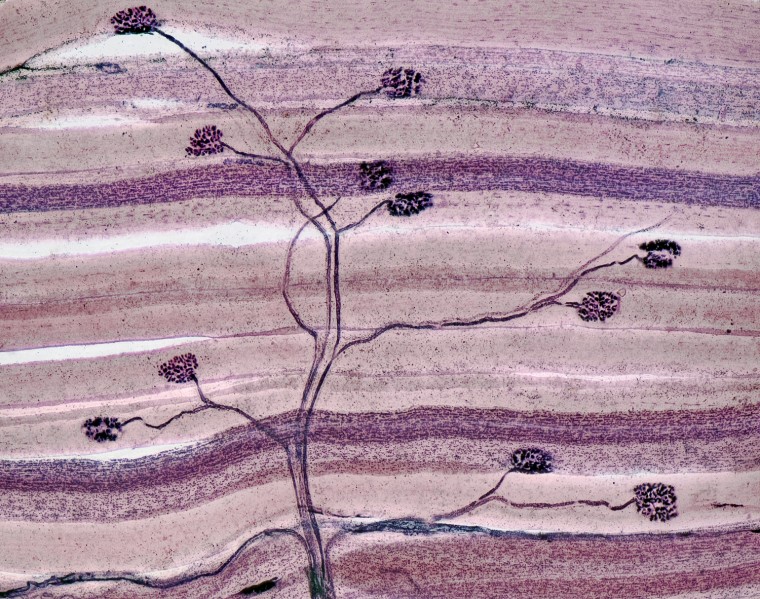
Nerve and muscle thin section
Brightfield, Image Stacking
40X
Axons of motor neurons synapsing with skeletal muscle cells

Tree of life

This 19th-place image by David Ward of Oakdale, Calif., shows a thin section of nerve and muscle cells at 40x magnification.

David G. Ward
Mr. Christian Sardet
Department of Life Sciences, Centre National de la Recherche Scientifique
Villefranche-sur-Mer, France
Annelid larva
Darkfield
100X

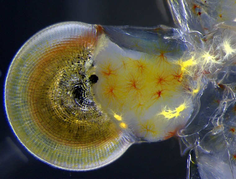
Early worm

This darkfield image by Christian Sardet of France's National Center for Scientific Research shows the larva of an annelid, or segmented worm, at 100x magnification. In darkfield microscopy, contrast is created by a bright specimen on a dark background. The image won 18th place in the Nikon Small World competition.
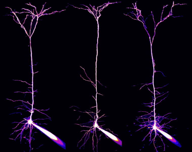
Dr. Alexandre William Moreau
Institute of Neurology, University College London
London, UK
Pyramidal neurons and their dendrites visualized in the visual cortex of a mouse brain
2-Photon, Focus Stacking, Fluorescence, Patch clamp
40X

Thinking about cheese

This 17th-place image by Alexandre William Moreau of University College London shows pyramidal neurons and their dendrites in the visual cortex of a mouse brain, seen at 40x magnification.

Mr. Geir Drange
Asker, Norway
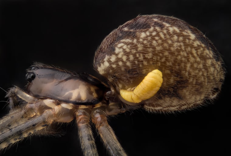
Pityohyphantes phrygianus (sheet weaver spider) with a parasitic wasp larva on the abdomen
Reflected Light, Focus Stacking
5X

Hitching a ride

This 16th-place image by Geir Drange of Asker, Norway, shows a parasitic wasp larva on the abdomen of a sheet weaver spider (Pityohyphantes phrygianus) at 5x magnification.

Dr. Christian Q. Scheckhuber
Goethe University
Frankfurt, Germany
Podospora anserina (fungus) filamentous tip cells
Fluorescence
630X

Glowing fungus

The Nikon Small World contest's 15th-place image was captured by Christian Q. Scheckhuber of Germany's Goethe University. It shows the filamentous tip cells of a type of fungus known as Podospora anserina at 630x magnification.

Mr. Zhong Hua
Department of Molecular Biology & Genetics, Johns Hopkins University School of Medicine
Baltimore, Maryland, USA
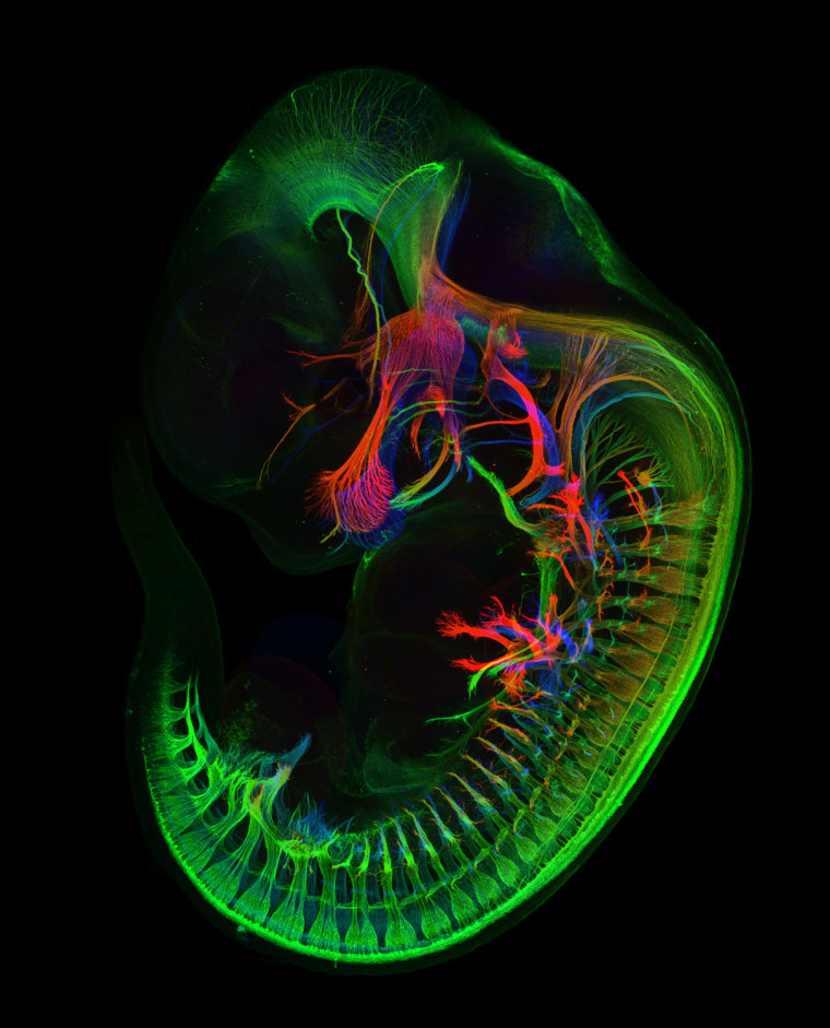
Peripheral nerves in E11.5 mouse embryo
Confocal
5X

Visible mouse

This 14th-place image by Zhong Hua of the Johns Hopkins University School of Medicine shows peripheral nerves in a mouse embryo at 5x magnification. Stains were used to highlight the different tissue types.

Dr. Michael Paul Nelson and Samantha Smith
Department of Pathology/Neuropathology, University of Alabama at Birmingham
Birmingham, Alabama, USA
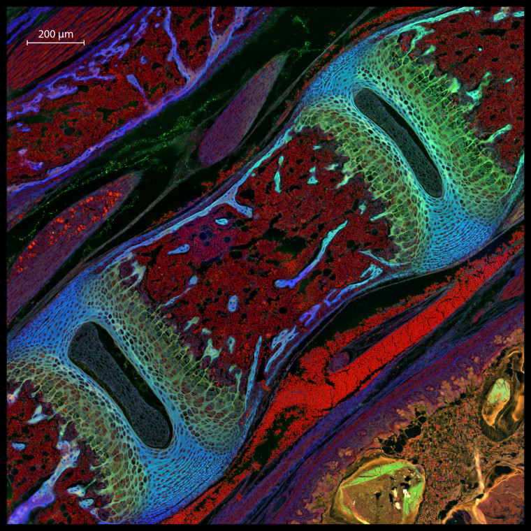
Mouse vertebra section
Focus Stacking
200X

Vertebra up close

This image by Michael Paul Nelson and Samantha Smith of the University of Alabama at Birmingham shows a vertebra section of a mouse at 200x.

Dr. Pedro Barrios-Perez
CPFC (nanofabrication), National Research Council of Canada/Information and Communication Technologies		
Ottawa, Ontario, Canada
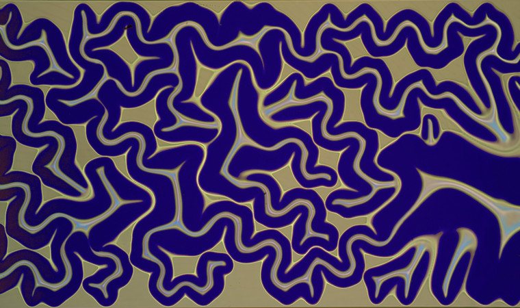
Silicon dioxide on polydimethylglutarimide-based resist
Bright field microscopy
200X

Study in silicon

This brightfield image by Pedro Barrios-Perez of the National Research Council of Canada shows silicon dioxide on polydimethylglutarimide-based resist at 200x magnification. The picture won 12th place in the Nikon Small World contest.

Miss Vitoria Tobias Santos
Universidade Federal do Rio de Janeiro, Rodrigo Evo Devo Group
Macaé, Rio de Janeiro, Brazil
Macrobrachium shrimp (ghost shrimp) eye
Stereomicroscopy
140X

Shrimp eyeball

Vitoria Tobias Santos of Brazil's Federal University of Rio de Janeiro captured this photo of the eye of a ghost shrimp (Macrobrachium) at 140x magnification. It won 11th place in the Nikon Small World contest.

Mr. Ted Kinsman
Department of Imaging and Photo Technology, Rochester Institute of Technology

Rochester, New York, USA
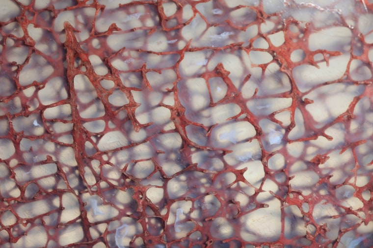
Thin section of a dinosaur bone preserved in clear agate
Focus Stacking
10X

Dino web

This 10th-place image by Ted Kinsman of the Rochester Institute of Technology shows a thin section of a dinosaur bone preserved in clear agate at 10x magnification.

Photographer:ted Kinsman
Mr. Mark A. Sanders
University Imaging Centers, University of Minnesota
Minneapolis, Minnesota, USA
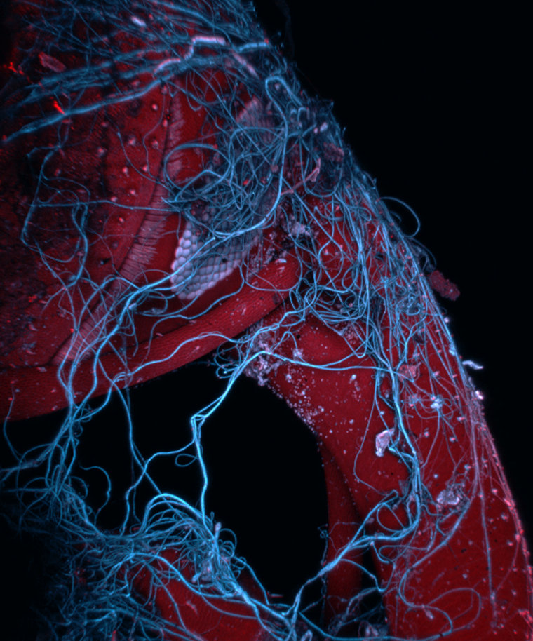
Insect wrapped in spider web
Confocal, Autofluorescence, Image Stacking
85X

Trapped

This ninth-place image by Mark A. Sanders of the University of Minnesota shows an insect wrapped in a spider web at 85x magnification.

Lilliputian leaf

This eighth-place image by Magdalena Turzańska of Poland's University of Wrocław shows a leafy liverwort, bryophyte plant and cyanobacteria at 50x magnification.

Dr. Jan Michels
Institute of Zoology, Functional Morphology and Biomechanics, Christian-Albrechts-Universität zu Kiel
Kiel, Germany
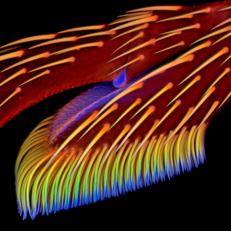
Adhesive pad on a foreleg of Coccinella septempunctata (ladybird beetle)
Confocal, Autofluorescence
20X

A beetle's brush

This seventh-place image by Jan Michels of Germany's University of Kiel shows the adhesive pad on the foreleg of a ladybird beetle (Coccinella septempunctata) at 20x magnification.

Miss Dorit Hockman
University of Cambridge
Cambridge, UK
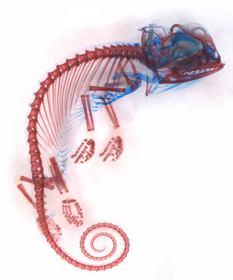
Chamaeleo calyptratus (veiled chameleon), embryo showing cartilage (blue) and bone (red)
Brightfield

Seeing through a chameleon

This sixth-place image by Dorit Hockman of the University of Cambridge shows the embryo of a veiled chameleon (Chamaeleo calyptratus). Cartilage is seen in blue, and bone in red.

Dr. Kieran Boyle
University of Glasgow, Institute of Neuroscience and Psychology
Glasgow, Scotland, UK
Hippocampal neuron receiving excitatory contacts
Fluorescence and Confocal
63X
[Improvision Data]

ImageName=
TimeStampMicroSeconds=3387346824918000
TimeStamp=10:40:24.918 on May 04, 2011
ChannelName=
ChannelNo=1
TimepointName=1
TimepointNo=1
ZPlane=1
BlackPoint=0
WhitePoint=255
WhiteColour=255,255,255
XCalibrationMicrons=0.154821
YCalibrationMicrons=0.154821
ZCalibrationMicrons=1
TotalChannels=1
TotalTimepoints=1
TotalZPlanes=1
Software=Volocity 4.4.0 Build 71
SampleUUID=bdab0a89-f1fd-466f-9fea-67a2c7e75f09

Feel the excitement

Kieran Boyle of the University of Glasgow, Scotland, captured this image of a hippocampal neuron receiving excitatory contacts at 63x magnification. The image came in fifth in the Nikon Small World contest.

Mr. Rogelio Moreno Gill
Panama City, Panamá
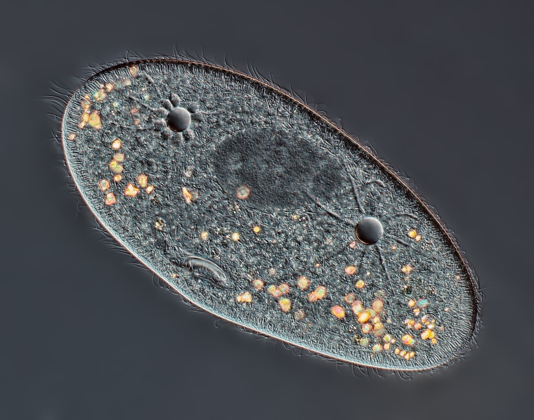
Paramecium sp. showing the nucleus, mouth and water expulsion vacuoles
Differential Interference Contrast
40X
Paramecium showing macronucleus, water expulsion vacuolas and mounth.  Nikon TE-300, plan apo 20x/0.75 objective, DIC, flash, Canon T3i mounted on the front port of the TE-300.

Panamanian paramecium

This fourth-place image by Rogelio Moreno Gill of Panama City, Panama, shows a paramecium, including the nucleus, mouth and water expulsion vacuoles at 40x magnification.

Rogelio Moreno Gill
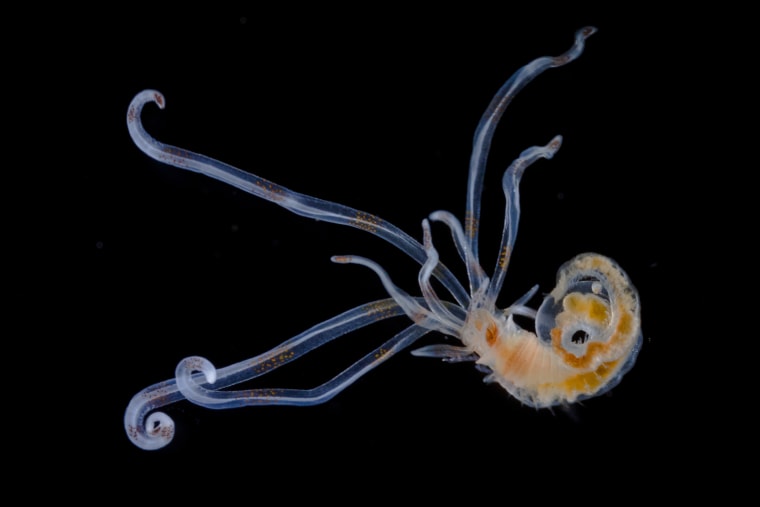
Dr. Alvaro Esteves Migotto
Universidade de São Paulo, Centro de Biologia Marinha
São Paulo, São Paulo, Brazil
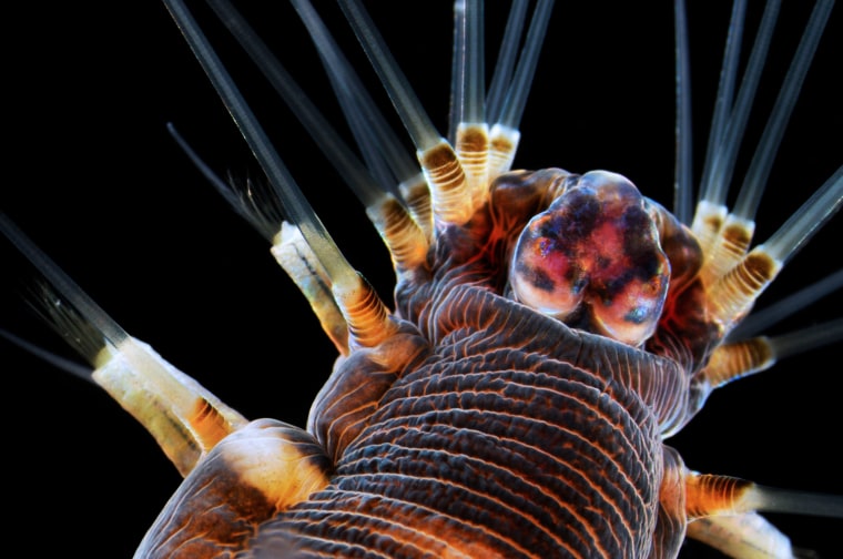
Marine worm
Stereomicroscopy, Darkfield
20X

Worm gone wild

This third-place image by Alvaro Esteves Migotto of the University of Sao Paulo shows a marine worm at 20x magnification.

Alvaro Migotto
Dr. Joseph Corbo
Washington University School of Medicine
St. Louis, Missouri, USA
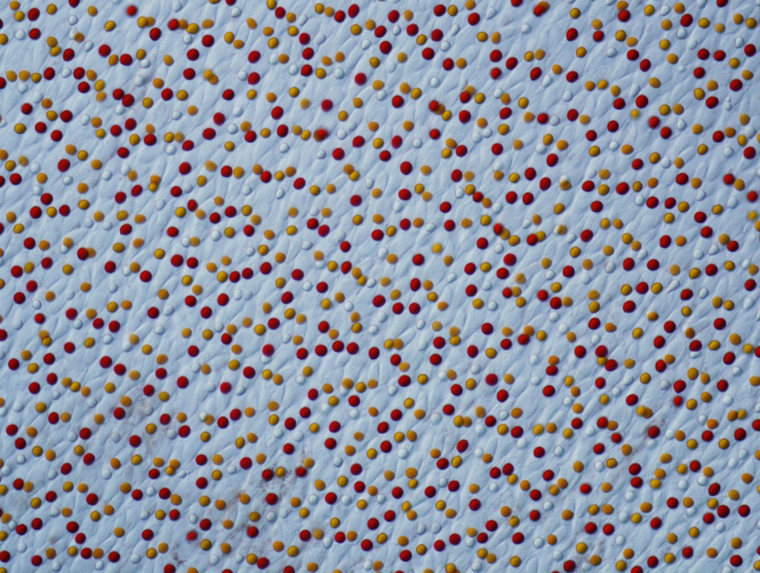
Chrysemys picta (painted turtle) retina
Differential Interference Contrast
400X

How a turtle sees

This second-place image by Joseph Corbo of the Washington University School of Medicine in St. Louis shows the retina of a painted turtle (Chrysemys picta) at 400x magnification.

Mr. Wim van Egmond
Micropolitan Museum
Berkel en Rodenrijs, Zuid Holland, The Netherlands
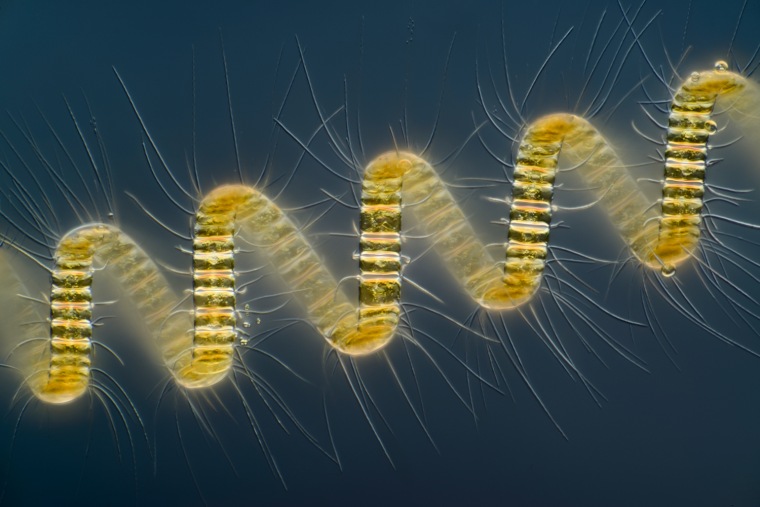
Chaetoceros debilis (marine diatom), a colonial plankton organism
Differential Interference Contrast, Image Stacking
250X

On a roll

Top honors in the 2013 Nikon Small World contest go to this image by Wim van Egmond of the Micropolitan Museum in the Netherlands. The image shows a marine diatom (Chaetoceros debilis), a colonial plankton organism, at 250x magnification.

1/20